Anatomy and Physiology of the Nose
Computer-based literature searches were used to identify all reports and reviews concerned with nasal conditions and SDB. These reports were read by the authors, reanalyzed, and grouped into those evaluating nasal-associated effects on SDB in normal control subjects, patients with nasal conditions or SDB, and children. 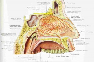
Anatomy and Physiology of the Nose
The nose is lined by pseudostratified epithelium resting on a basement membrane, separating it from deeper submucosal layers. The submucosa contains mucous, seromucous, and serous glands. The small arteries, arterioles, and arteriovenous anastamoses determine regional blood flow. Capacitance vessels, consisting of veins and cavernous sinusoids, determine nasal patency. Constriction and relaxation of these venous capacitance vessels is regulated by the sympathetic nervous system Viagra online Australia. The cavernous sinusoids lie beneath the capillaries and venules, are most dense in the inferior and middle turbinates, and contain smooth-muscle cells controlled by the sympathetic nervous system. Loss of sympathetic tone or, to a lesser degree, cholinergic stimulation causes this sinusoidal erectile tissue to become engorged. Cholinergic stimulation causes arterial dilation and promotes the passive diffusion of plasma proteins into glands and the active secretion by mucous glands in cells.
Novel neurotransmitters, including substance P, calcitonin gene-related peptide, and vasointestinal peptide, have been detected in nasal secretions after nasal allergen challenge of patients with allergic rhinitis. Antidromic stimulation of sensory nerve fibers in the nose can release a variety of neurotransmitters including substance P, a mediator of increased vascular permeability. Because neurotransmitters also produce changes in regional blood flow and glandular secretion, their role in rhinitis may be important.